Loculated Pleural Effusion Diagram : Chest Drainage Abstract Europe Pmc - Surgical treatment of pleural effusion may include chest.. We studied the value of transca … Light the author22 has estimated that more than 1 million cases of pleural effusion occur annually in the united states. This condition involves the buildup of fluid between the tissues (pleura) that line the lungs and the inside of the chest wall. Surgical treatment of pleural effusion may include chest. Loculated effusions are difficult to confirm with chest radiograph, but ultrasound, computed tomography (ct), and even magnetic resonance imaging (mri) may be used to verify a localized collection of pleural fluid.
The first step in the evaluation of a pleural effusion is to determine whether the pleural fluid is a transudate or an exudate. The reasons for effusion are many, and the specific diagnosis is often based upon tap or drainage of the fluid. Diagnosed stage 4 nsclc 1 year ago, had massive pleural effusions that needed to be drained every 3 weeks, had the right side talced but it was so painful that i didn't want to do that on the left. Pleural effusions in the intensive care setting. Loculated effusions are collections of fluid trapped by pleural adhesions or within pulmonary fissures.
Http Indiachest Org Wp Content Uploads 2016 07 Intrapleural Agents For Pleural Infections Nagarjun 2013 Pdf from Diuretics and other heart failure medications are used to treat pleural effusion caused by congestive heart failure or other medical causes. The first step in the evaluation of a pleural effusion is to determine whether the pleural fluid is a transudate or an exudate. Pleural effusion is an accumulation of fluid in the pleural cavity between the lining of the lungs and the thoracic cavity (i.e., the visceral and parietal for recurrent pleural effusion or urgent drainage of infected and/or loculated effusions 2728. Diagnosed stage 4 nsclc 1 year ago, had massive pleural effusions that needed to be drained every 3 weeks, had the right side talced but it was so painful that i didn't want to do that on the left. Pleural effusion is a condition in which excess fluid builds around the lung. Loculated effusions, defined as effusions that do not shift freely in the pleural space, occur when there are adhesions between the visceral and parietal pleura. Previous studies have demonstrated that intrapleural administration of tissue plasminogen activator (tpa) combined with human recombinant dnase can improve fluid drainage and reduce surgery for patients with loculated parapneumonic effusions; Streptokinase appears to improve the resolution of loculated pleural effusions when chest tube drainage fails to achieve symptomatic relief.
What are the different appearances of pleural effusion? Malignant pleural effusion is a frequent complication of some common cancers. Surgical thoracostomy tube placement and radiologically guided catheter drainage are standard therapy for loculated pleural fluid collections. Transudative effusions are a result of pressure filtration without capillary injury (i.e hydrostatic and oncotic pressure abnormalities). Most effusions start like this and can be easily missed. Treatment of pleural effusion is based on the underlying condition and whether the effusion is causing severe respiratory symptoms, such as shortness of breath or difficulty breathing. The first step in the evaluation of a pleural effusion is to determine whether the pleural fluid is a transudate or an exudate. A pleural effusion is accumulation of excessive fluid in the pleural space, the potential space that surrounds each lung.under normal conditions, pleural fluid is secreted by the parietal pleural capillaries at a rate of 0.01 millilitre per kilogram weight per hour, and is cleared by lymphatic absorption leaving. However, the efficacy of this treatment has not been evaluated for complicated pleural effusions. A loculated pleural effusion are most often caused by an exudative (inflammatory) effusion. Pleural effusions in the intensive care setting. Diagram of fluid buildup in the pleura: Loculated pleural effusion ct scan.ct scan of the chest of a patient with large loculated pleural effusion in his left thoracic cavity.
Pleural effusion is extra fluid around the lung. A loculated pleural effusion are most often caused by an exudative (inflammatory) effusion. Prior chest radiographs indicating that the blunting is a new finding also provide a good indicator of pleural effusion. The first step in the evaluation of a pleural effusion is to determine whether the pleural fluid is a transudate or an exudate. Loculated pleural effusion ct scan.ct scan of the chest of a patient with large loculated pleural effusion in his left thoracic cavity.
Imaging Findings Of Free Flowing Effusion Online Medical Library from d3uigcfkiiww0g.cloudfront.net Loculated right pleural effusion with foci of atelectasis and consolidative changes concerning for pneumonia. Loculated effusions are difficult to confirm with chest radiograph, but ultrasound, computed tomography (ct), and even magnetic resonance imaging (mri) may be used to verify a localized collection of pleural fluid. Most malignant effusions can be controlled by thoracentesis and/or closed thoracostomy tube drainage and sclerosis of the pleural cavity. Loculated malignant effusions however, are inherently resistant to the usual approaches because of nonexpanding underlying lung. Prior chest radiographs indicating that the blunting is a new finding also provide a good indicator of pleural effusion. The formation of a transudate usually results from increased capillary hydrostatic pressure or from decreased colloid osmotic pressure. Causes of an exudative effusion are malignancy, infection, or inflammatory disorders such as rheumatoid arthritis. The first step in the evaluation of a pleural effusion is to determine whether the pleural fluid is a transudate or an exudate.
In vitro efficacy of varidase versus streptokinase or urokinase for liquefying thick purulent exudative material from loculated empyema.
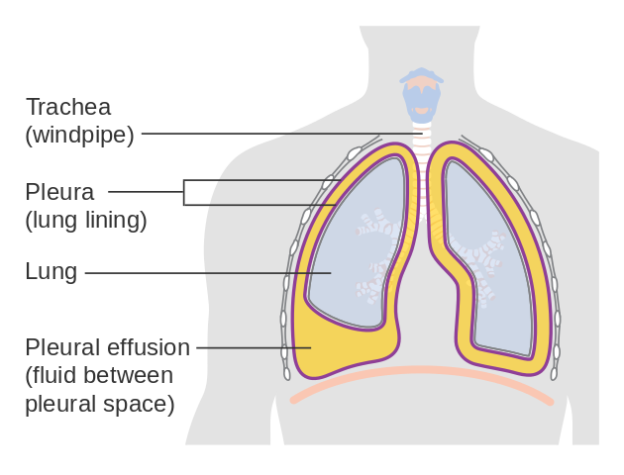
Pleura l effusion seen in an ultra sound image as in one or more fixed pockets in the pleural space is said to be loculated pleural effusion.in us scan us scan they can be identified clearly and it is very complicated.pleural effusion generally found the space. The first step in the evaluation of a pleural effusion is to determine whether the pleural fluid is a transudate or an exudate. Various types of pneumonia, a lung infection, can cause atelectasis. Most effusions start like this and can be easily missed. Loculated pleural effusion ct scan.ct scan of the chest of a patient with large loculated pleural effusion in his left thoracic cavity. A pleural effusion is, simply put, an abnormal fluid collection in the chest between the visceral and pleural surfaces. Treatment of pleural effusion is based on the underlying condition and whether the effusion is causing severe respiratory symptoms, such as shortness of breath or difficulty breathing. In left heart failure, which results in elevated pressures in the venous system, there is usually some accumulation of fluid in the pleural space. Diagram of fluid buildup in the pleura: The etiologies of pleural effusions as a whole, and then more specifically the various specific findings of pleural effusions resulting from infectious diseases, will be examined. Causes of an exudative effusion are malignancy, infection, or inflammatory disorders such as rheumatoid arthritis. Pleural effusion is extra fluid around the lung. This condition involves the buildup of fluid between the tissues (pleura) that line the lungs and the inside of the chest wall.
Prior chest radiographs indicating that the blunting is a new finding also provide a good indicator of pleural effusion. Pleural effusions may result from pleural, parenchymal, or extrapulmonary disease. Pleural effusion is extra fluid around the lung. Light the author22 has estimated that more than 1 million cases of pleural effusion occur annually in the united states. The possibility of a pleural effusion should be considered whenever a patient with an abnormal chest radiograph is evaluated.
Parapneumonic Pleural Effusions And Empyema Thoracis Article from www.statpearls.com Loculated malignant effusions however, are inherently resistant to the usual approaches because of nonexpanding underlying lung. However, the efficacy of this treatment has not been evaluated for complicated pleural effusions. Terminology pleural effusion is commonly used as. Icu patients cannot sit up and the effusion layers posteriorly. Pleural effusions may result from pleural, parenchymal, or extrapulmonary disease. The formation of a transudate usually results from increased capillary hydrostatic pressure or from decreased colloid osmotic pressure. Light the author22 has estimated that more than 1 million cases of pleural effusion occur annually in the united states. Pleural effusions in the intensive care setting.
Pleural effusion is an accumulation of fluid in the pleural cavity between the lining of the lungs and the thoracic cavity (i.e., the visceral and parietal for recurrent pleural effusion or urgent drainage of infected and/or loculated effusions 2728.
Transudative effusions are a result of pressure filtration without capillary injury (i.e hydrostatic and oncotic pressure abnormalities). It didn't work totally on the right but left me with about 500 ml of loculated fluid, and it did stop the effusions on the right side. Previous studies have demonstrated that intrapleural administration of tissue plasminogen activator (tpa) combined with human recombinant dnase can improve fluid drainage and reduce surgery for patients with loculated parapneumonic effusions; Etiology, prevalence, and epidemiology pleural effusion is the accumulation of fluid in the pleural space resulting from disruption of the homeostatic forces responsible for the movement of pleural fluid. Most effusions start like this and can be easily missed. Prior chest radiographs indicating that the blunting is a new finding also provide a good indicator of pleural effusion. Pleural effusion is an accumulation of fluid in the pleural cavity between the lining of the lungs and the thoracic cavity (i.e., the visceral and parietal for recurrent pleural effusion or urgent drainage of infected and/or loculated effusions 2728. Streptokinase appears to improve the resolution of loculated pleural effusions when chest tube drainage fails to achieve symptomatic relief. Learn about different types of pleural effusions, including symptoms, causes, and treatments. Pleural effusions in the intensive care setting. Icu patients cannot sit up and the effusion layers posteriorly. Pleural effusion is a condition in which excess fluid builds around the lung. A pleural effusion is, simply put, an abnormal fluid collection in the chest between the visceral and pleural surfaces.
The etiologies of pleural effusions as a whole, and then more specifically the various specific findings of pleural effusions resulting from infectious diseases, will be examined loculated pleural effusion. The etiologies of pleural effusions as a whole, and then more specifically the various specific findings of pleural effusions resulting from infectious diseases, will be examined.
0 Comments:
Posting Komentar