Home
Uncategories
Anatomy Of Chest X Ray - Chest X Ray Mediastinum Anatomy My Own Rad - The interpretation of a chest film requires the understanding of basic principles.
Anatomy Of Chest X Ray - Chest X Ray Mediastinum Anatomy My Own Rad - The interpretation of a chest film requires the understanding of basic principles.
Anatomy Of Chest X Ray - Chest X Ray Mediastinum Anatomy My Own Rad - The interpretation of a chest film requires the understanding of basic principles.. Normal chest x ray radiological anatomy is where your human anatomy knowledge meets clinical practice. This noninvasive method produces images of the heart, lungs, airways, blood vessels, and the bones of the chest and spine(1). Select questions from the bottom options. In this article we will focus on: Because some conditions of the chest.
In fact every radiologst should be an expert in chest film reading. The chest or thorax is the region between the neck and diaphragm that encloses organs, such as the heart, lungs, esophagus, trachea, and thoracic diaphragm. Use the mouse to select the site and click. The trachea passes to the right of the aorta and so may be slightly off midline to the right. This noninvasive method produces images of the heart, lungs, airways, blood vessels, and the bones of the chest and spine(1).
The Chest X Ray A Systematic Teaching Atlas Matthias Hofer 9781588905543 Amazon Com Books from images-na.ssl-images-amazon.com There is much deeper anatomy than that and maybe we will touch on segmental airway anatomy when we tackle ct of the chest, but for now this is definitely enough. Because some conditions of the chest. Other important structures, such as the pleura, only become visible when abnormal, and some are not visible at all, such as the phrenic nerve. Chest radiographs are the most common film taken in medicine. Adapted here for independent study. This noninvasive method produces images of the heart, lungs, airways, blood vessels, and the bones of the chest and spine(1). Normal anatomy the pleural cavity is formed by the visceral pleura (= membrane attached to the lungs) and the parietal pleura (= membrane attached to the surrounding structures). Anatomy of chest x ray.
17 on page 22, fig.
The sternum is also included on a frontal view but it overlies other midline structures and so is obscured. The interpretation of a chest film requires the understanding of basic principles. Chest radiographs are the most common film taken in medicine. Posted by radiologypics ⋅ march 17, 2013 ⋅ leave a comment. Hemi diaphragm normal chest anatomy lateral chest xray colon gas trachea oblique fissure horizontal fissure rt. Yes, it is the same patient 2. Filed under anatomy, chest, chest radiograph. Anatomy of chest x ray. The bones are used as useful markers of chest radiograph quality. The chest films selected represent commonly occurring cardiopulmonary problems in the primary care setting Doctors use them to diagnose problems. Normal anatomy the pleural cavity is formed by the visceral pleura (= membrane attached to the lungs) and the parietal pleura (= membrane attached to the surrounding structures). Identify the location of carina and click 1.
L the portion of the left lung that corresponds anatomically to the right middle lobe is incorporated into the left upper lobe. Chest radiographs are the most common film taken in medicine. Normal chest x ray radiological anatomy is where your human anatomy knowledge meets clinical practice. The chest films selected represent commonly occurring cardiopulmonary problems in the primary care setting They contain air and so are of lower density (blacker) than the surrounding soft tissues.
Chest X Ray Interpretation Ppt Video Online Download from slideplayer.com Posted by radiologypics ⋅ march 17, 2013 ⋅ leave a comment. Yes, it is the same patient 2. Doctors use them to diagnose problems. You may also find aortic arch, pulmonary trunk, left main bronchus, left atrium, left ventricle, oblique fissure, diaphragm, gastric bubble as well. This noninvasive method produces images of the heart, lungs, airways, blood vessels, and the bones of the chest and spine(1). Normal chest x ray radiological anatomy is where your human anatomy knowledge meets clinical practice. Anatomy of chest x ray. Adapted here for independent study.
Normal chest x ray radiological anatomy is where your human anatomy knowledge meets clinical practice.
Basics of chest x ray. Posted by radiologypics ⋅ march 17, 2013 ⋅ leave a comment. There is much deeper anatomy than that and maybe we will touch on segmental airway anatomy when we tackle ct of the chest, but for now this is definitely enough. Identify the location of carina and click 1. Because some conditions of the chest. Computed tomography (ct) of the chest can detect pathology that may not show up on a conventional chest radiograph (1). A line is drawn from anterior surface of the body of 6th thoracic vertebrae passing through the apex of the heart up to anterior lower most part of diaphragm. You may also find aortic arch, pulmonary trunk, left main bronchus, left atrium, left ventricle, oblique fissure, diaphragm, gastric bubble as well. They contain air and so are of lower density (blacker) than the surrounding soft tissues. No, it is not the same patient Doctors use them to diagnose problems. The sternum is also included on a frontal view but it overlies other midline structures and so is obscured. When the heart is located in front of the column, and the aortic arch in not visible, this is a right anterior oblique (fig.
Normal chest x ray radiological anatomy is where your human anatomy knowledge meets clinical practice. The bones are used as useful markers of chest radiograph quality. Anatomically, the heart is located in the anterior thoracic cavity; Other important structures, such as the pleura, only become visible when abnormal, and some are not visible at all, such as the phrenic nerve. Normal anatomy the pleural cavity is formed by the visceral pleura (= membrane attached to the lungs) and the parietal pleura (= membrane attached to the surrounding structures).
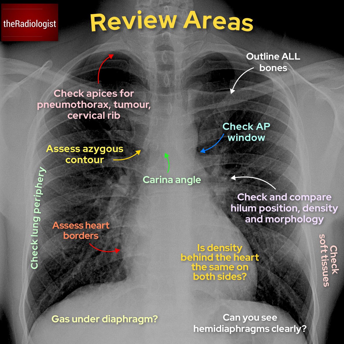
Theradiologist On Twitter Basics Chest X Ray Anatomy And Review Areas Foamrad Foamed from pbs.twimg.com Identify the location of carina and click 1. Normal anatomy the pleural cavity is formed by the visceral pleura (= membrane attached to the lungs) and the parietal pleura (= membrane attached to the surrounding structures). Basics of chest x ray. The trachea passes to the right of the aorta and so may be slightly off midline to the right. This noninvasive method produces images of the heart, lungs, airways, blood vessels, and the bones of the chest and spine(1). Yes, it is the same patient 2. Anatomy of chest x ray. Doctors use them to diagnose problems.
The interpretation of a chest film requires the understanding of basic principles.
In this article we will focus on: Posted by radiologypics ⋅ march 17, 2013 ⋅ leave a comment. Doctors use them to diagnose problems. Chest radiographs are the most common film taken in medicine. Anatomy of chest x ray. Identify the location of carina and click 1. The trachea passes to the right of the aorta and so may be slightly off midline to the right. Other important structures, such as the pleura, only become visible when abnormal, and some are not visible at all, such as the phrenic nerve. The chest films selected represent commonly occurring cardiopulmonary problems in the primary care setting Adapted here for independent study. 17 on page 22, fig. Computed tomography (ct) of the chest can detect pathology that may not show up on a conventional chest radiograph (1). No, it is not the same patient
The bones are used as useful markers of chest radiograph quality anatomy of chest. Normal chest x ray radiological anatomy is where your human anatomy knowledge meets clinical practice.


0 Comments:
Posting Komentar